

GIVE THE PERFECT GIFT
Erin Mills Town Centre Gift Cards are the perfect choice for your gift giving needs.Purchase gift cards at kiosks near the food court or centre court, at Guest Services, or click below to purchase online.PURCHASE HEREHome
Keratoconjunctivitis Sicca and Filamentary Keratopathy: Vivo Morphology the Human Cornea Conjunctiva
Indigo
Loading Inventory...
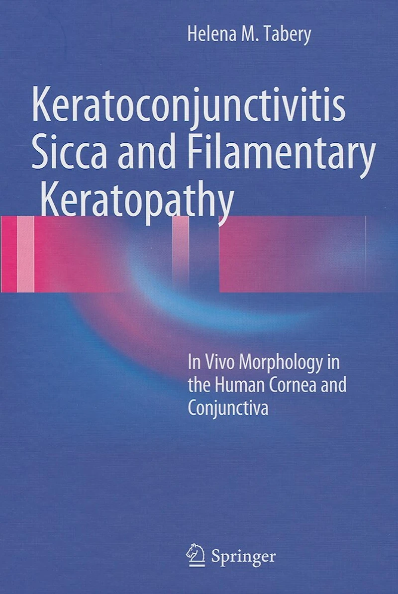
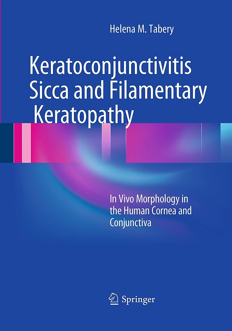
Keratoconjunctivitis Sicca and Filamentary Keratopathy: Vivo Morphology the Human Cornea Conjunctiva
Current price: $248.50

Keratoconjunctivitis Sicca and Filamentary Keratopathy: Vivo Morphology the Human Cornea Conjunctiva
Current price: $248.50
Loading Inventory...
Size: Hardcover
*Product information may vary - to confirm product availability, pricing, shipping and return information please contact Indigo
This book presents in vivo captured high-magnification images of two conditions: keratoconjunctivitis sicca (KCS, or dry eye), an extremely common disease of the ocular surface, and filamentary keratopathy, a relatively rare phenomenon most commonly associated with KCS. The images of KCS represent the broad spectrum of ocular surface changes seen in the condition while the images of filamentary keratopathy clearly reveal the components of the ocular surface appendices, termed filaments. The photographs show phenomena captured in various illumination modes, without staining and after staining with diagnostic dyes, and the photographic sequences illustrate their dynamics. The images reflect the in vivo situation. Once aware of the various phenomena, anyone working with standard diagnostic equipment - the slit lamp and the diagnostic dyes- will be able to detect almost all of them. The book will be invaluable for all who deal with ocular surface diseases.
This book presents in vivo captured high-magnification images of two conditions: keratoconjunctivitis sicca (KCS, or dry eye), an extremely common disease of the ocular surface, and filamentary keratopathy, a relatively rare phenomenon most commonly associated with KCS. The images of KCS represent the broad spectrum of ocular surface changes seen in the condition while the images of filamentary keratopathy clearly reveal the components of the ocular surface appendices, termed filaments. The photographs show phenomena captured in various illumination modes, without staining and after staining with diagnostic dyes, and the photographic sequences illustrate their dynamics. The images reflect the in vivo situation. Once aware of the various phenomena, anyone working with standard diagnostic equipment - the slit lamp and the diagnostic dyes- will be able to detect almost all of them. The book will be invaluable for all who deal with ocular surface diseases.
You May Also Like












More About Indigo at Erin Mills Town Centre
The largest book retailer in Canada also offers toys, music, home décor, gifts and lifestyle products. What's Inside...Books, Magazines, CD’s and DVD’s, Toys and Gifts, Home Accents, Electronics, Baby’s and Children’s Section, Bath and Body, Kitchen and Bedroom, Stationary Located outside in the exterior plaza.
5015 Glen Erin Dr, Mississauga, ON L5M 0R7, Canada
Find Indigo at Erin Mills Town Centre in Mississauga ON
Visit Indigo at Erin Mills Town Centre in Mississauga ON